Normal brain (MRI) Radiology Case Radiopaedia.org Brain MRI. Brain MRI Indications : 1- Multiple sclerosis MS 2- Primary tumor assessment and metastasis. 3- evaluation of infarction. 4- Unexplained neurological symptoms and defect Brain MRI Equipment : 1- Head coil (circularly polarized (CP) head coils. These volume coils …
Brain Atlas of human anatomy with MRI
MRI In Practice Download Free (EPUB PDF). 5/17/2018 · Rapid-sequence MRI of the brain (also known as “ultrafast brain,” “quick brain,” “fast brain,” and “one bang” MRI) has long been used in the evaluation of ventricular shunt catheters due to its ability to quickly evaluate intracranial fluid–containing spaces without anesthesia or the ionizing radiation of CT., Updating… Please wait. Unable to process the form. Check for errors and try again..
Similar to CT a systematic approach is best when interpreting MRI brain. MRIs of the brain can be intimidating at first sight because of all the different sequences and parameters. However, the same general principles of CT head interpretation apply, as the first steps are anatomically-based after all. Similar to CT a systematic approach is best when interpreting MRI brain. MRIs of the brain can be intimidating at first sight because of all the different sequences and parameters. However, the same general principles of CT head interpretation apply, as the first steps are anatomically-based after all.
4/21/2019 · You need data from MRI tomography, a volumetric modeling that scans 2D slices of a volume and combines these into one 3D data set. There are various software applications that can translate such data to images, and even export the data as a 3D fil... Brain MRI. Brain MRI Indications : 1- Multiple sclerosis MS 2- Primary tumor assessment and metastasis. 3- evaluation of infarction. 4- Unexplained neurological symptoms and defect Brain MRI Equipment : 1- Head coil (circularly polarized (CP) head coils. These volume coils …
! 5!! The!evaluation!of!brain!volume!is!divided!global!and!regional!components.!Whereas!global! assessment!of!volumeis!based!on!ageneral!assessment!of!theprominenceof pdf. A new Method on Brain MRI Image Preprocessing for Tumor Detection Engineering and Technology A new Method on Brain MRI Image Preprocessing for Tumor Detection D. Arun Kumar Department of ECE, GMRIT RAJAM, AP, INDIA ABSTRACT Medical imaging is a vital component of a large number of applications. MRI is the state-of the-art medical
11/6/2019 · The basic types of sequences used in brain MRI create either T1-weighted or T2-weighted images. In T1-weighted images, CSF and fluid appear dark. Gray matter is darker than white matter. Patients are instructed to lie supine and to stay still with their … MRI of the head: Magnetic resonance imaging (MRI) uses a powerful magnetic field, radio frequency pulses and a computer to produce detailed pictures of organs, soft tissues, bone and virtually all other internal body structures. MRI provides detailed images that can detect brain abnormalities such as tumors and infection.
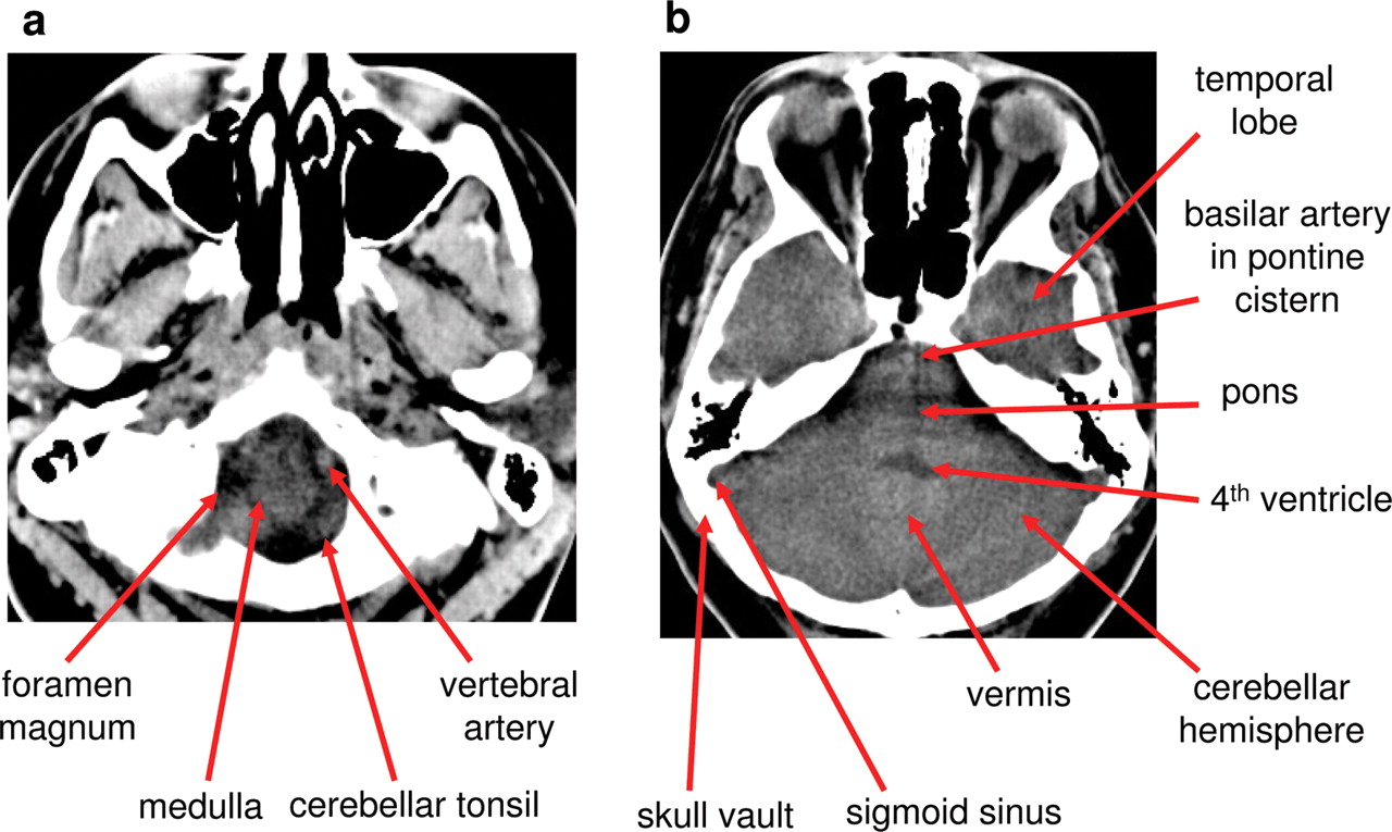
Brain MRI. Brain MRI Indications : 1- Multiple sclerosis MS 2- Primary tumor assessment and metastasis. 3- evaluation of infarction. 4- Unexplained neurological symptoms and defect Brain MRI Equipment : 1- Head coil (circularly polarized (CP) head coils. These volume coils … MRI Atlas of the Brain. This page presents a comprehensive series of labeled axial, sagittal and coronal images from a normal human brain magnetic resonance imaging exam. This MRI brain cross-sectional anatomy tool serves as a reference atlas to guide radiologists and researchers in the accurate identification of the brain structures.
7/27/2017 · These MRI images can show the existence of brain tumors and their growth being experienced, blood vessel blockage in the brain (and the severity), as well as other signs of disease. But in order to determine what an abnormal brain MRI looks like, a medical professional must first know what a normal one looks like. Updating… Please wait. Unable to process the form. Check for errors and try again.
8/1/2019В В· BACKGROUND AND PURPOSE: Classic findings of intracranial hypotension on MR imaging, such as brain stem slumping, can be variably present and, at times, subjective, potentially making the diagnosis difficult. We hypothesize that the angle between the cerebral peduncles correlates with the volume of interpeduncular cistern fluid and is decreased in cases of intracranial hypotension. Department of Engineering Science Oxford University Michaelmas 2004. MRI is used for a huge range of clinical applications Gaussian pdf for GM and that for WM This means that there are many misclassifications of Work done in the Functional MRI of the Brain Laboratory by Tim Behrens, Dr. Johansson-Berg, Mike Brady, Paul Matthews, Steve
3/9/2014В В· Read Knee MRI for ACL and PCL. A patient's guide to reading your own knee MRI for possible Anterior Cruciate Ligament (ACL) or Posterior Cruciate Ligament (PCL) injury. 11/11/2013В В· General practitioners (GPs) are expected to be allowed to request MRI scans for adults for selected clinically appropriate indications from November 2013 as part of the expansion of Medicare-funded MRI services announced by the Federal Government in 2011. 1 This article aims to give a brief overview of MRI brain imaging relevant to GPs, which will facilitate explanation of scan findings and
Department of Engineering Science Oxford University Michaelmas 2004. MRI is used for a huge range of clinical applications Gaussian pdf for GM and that for WM This means that there are many misclassifications of Work done in the Functional MRI of the Brain Laboratory by Tim Behrens, Dr. Johansson-Berg, Mike Brady, Paul Matthews, Steve Brain MRI. Brain MRI Indications : 1- Multiple sclerosis MS 2- Primary tumor assessment and metastasis. 3- evaluation of infarction. 4- Unexplained neurological symptoms and defect Brain MRI Equipment : 1- Head coil (circularly polarized (CP) head coils. These volume coils …
11/6/2019 · The basic types of sequences used in brain MRI create either T1-weighted or T2-weighted images. In T1-weighted images, CSF and fluid appear dark. Gray matter is darker than white matter. Patients are instructed to lie supine and to stay still with their … Similar to CT a systematic approach is best when interpreting MRI brain. MRIs of the brain can be intimidating at first sight because of all the different sequences and parameters. However, the same general principles of CT head interpretation apply, as the first steps are anatomically-based after all.
Brain Tumor MRI Magnetic Resonance Imaging Image

(PDF) A machine learning technique for MRI brain images. Updating… Please wait. Unable to process the form. Check for errors and try again., Department of Engineering Science Oxford University Michaelmas 2004. MRI is used for a huge range of clinical applications Gaussian pdf for GM and that for WM This means that there are many misclassifications of Work done in the Functional MRI of the Brain Laboratory by Tim Behrens, Dr. Johansson-Berg, Mike Brady, Paul Matthews, Steve.
Head MRI Purpose Preparation and Procedure

Brain Tumors RadiologyInfo.org. Brain Tumor Treatment Brain Tumors Overview A brain tumor is a group of abnormal cells that grows in or around the brain. Tumors can directly destroy healthy brain cells. They can also indirectly damage healthy cells by crowding other parts of the brain and causing … https://nl.wikipedia.org/wiki/MRI-scanner Similar to CT a systematic approach is best when interpreting MRI brain. MRIs of the brain can be intimidating at first sight because of all the different sequences and parameters. However, the same general principles of CT head interpretation apply, as the first steps are anatomically-based after all..

3/9/2014В В· Read Knee MRI for ACL and PCL. A patient's guide to reading your own knee MRI for possible Anterior Cruciate Ligament (ACL) or Posterior Cruciate Ligament (PCL) injury. 7/13/2016В В· Brain Imaging With MRI And CT [ PDF] [ United VRG] Topics tamer karam Collection opensource Language English. ark:/13960/t3421r584 Ocr ABBYY FineReader 11.0 Ppi 600 Scanner Internet Archive HTML5 Uploader 1.6.3. plus-circle Add Review. comment. Reviews There are no reviews yet. Be the first one to write a review. 4,804 Views PDF download.
Magnetic resonance imaging (MRI) of the head is a painless, noninvasive test that produces detailed images of your brain and brain stem. An MRI machine creates the images using a magnetic field ! 5!! The!evaluation!of!brain!volume!is!divided!global!and!regional!components.!Whereas!global! assessment!of!volumeis!based!on!ageneral!assessment!of!theprominenceof
pdf. A new Method on Brain MRI Image Preprocessing for Tumor Detection Engineering and Technology A new Method on Brain MRI Image Preprocessing for Tumor Detection D. Arun Kumar Department of ECE, GMRIT RAJAM, AP, INDIA ABSTRACT Medical imaging is a vital component of a large number of applications. MRI is the state-of the-art medical An MRI (magnetic resonance imaging) lets your doctor see the organs, bones, and tissues inside your body without having to do surgery. This test can help diagnose a disease or injury. You might
1MRI Brain W W/O Contrast 70553 Contrast - Knee, Ankle, Mid/Hindfoot, Hip Contrast - Shoulder, Elbow, Wrist #MRI Spine Cervical W W/O Contrast 72156 1MRI Breast W/O Contrast 77047 1MRI Extremity Lower Joint W W/O 73723 1MR Enterography 74183, 72197 #MRI Spine Lumbar W/O Contrast 72148 1/1/2017В В· Objective Many neonatal intensive care units (NICUs) have adopted the practice of performing routine brain MRI in very low birth weight (VLBW) infants at term-equivalent age in order to better evaluate prematurity-related acquired lesions. A number of unexpected brain abnormalities of potential clinical significance can be visualised on routine scans as well.
Brain Tumor MRI - Free download as Powerpoint Presentation (.ppt / .pptx), PDF File (.pdf), Text File (.txt) or view presentation slides online. this is a project proposal presentation explaining the detection of tumors in the brain from the analysis of brain MRI images. the project is to be implemented using the MATLAB programming environment. Similar to CT a systematic approach is best when interpreting MRI brain. MRIs of the brain can be intimidating at first sight because of all the different sequences and parameters. However, the same general principles of CT head interpretation apply, as the first steps are anatomically-based after all.
3/9/2014В В· Read Knee MRI for ACL and PCL. A patient's guide to reading your own knee MRI for possible Anterior Cruciate Ligament (ACL) or Posterior Cruciate Ligament (PCL) injury. 1MRI Brain W W/O Contrast 70553 Contrast - Knee, Ankle, Mid/Hindfoot, Hip Contrast - Shoulder, Elbow, Wrist #MRI Spine Cervical W W/O Contrast 72156 1MRI Breast W/O Contrast 77047 1MRI Extremity Lower Joint W W/O 73723 1MR Enterography 74183, 72197 #MRI Spine Lumbar W/O Contrast 72148
Brain Tumor Treatment Brain Tumors Overview A brain tumor is a group of abnormal cells that grows in or around the brain. Tumors can directly destroy healthy brain cells. They can also indirectly damage healthy cells by crowding other parts of the brain and causing … MRI Atlas of the Brain. This page presents a comprehensive series of labeled axial, sagittal and coronal images from a normal human brain magnetic resonance imaging exam. This MRI brain cross-sectional anatomy tool serves as a reference atlas to guide radiologists and researchers in the accurate identification of the brain structures.
Magnetic resonance imaging (MRI) of the head is a painless, noninvasive test that produces detailed images of your brain and brain stem. An MRI machine creates the images using a magnetic field 11/11/2013В В· General practitioners (GPs) are expected to be allowed to request MRI scans for adults for selected clinically appropriate indications from November 2013 as part of the expansion of Medicare-funded MRI services announced by the Federal Government in 2011. 1 This article aims to give a brief overview of MRI brain imaging relevant to GPs, which will facilitate explanation of scan findings and
Magnetic resonance imaging (MRI) is a medical imaging technique used in radiology to form pictures of the anatomy and the physiological processes of the body. MRI scanners use strong magnetic fields, magnetic field gradients, and radio waves to generate images of the organs in the body. the brain. In this T1-weighted image, grey matter is lightly coloured, while white matter • MRI scans require patients to hold very still for long periods of time up to 90 minutes or more in some cases Illustration of the MR read gradient and signals generated at different
pdf. A new Method on Brain MRI Image Preprocessing for Tumor Detection Engineering and Technology A new Method on Brain MRI Image Preprocessing for Tumor Detection D. Arun Kumar Department of ECE, GMRIT RAJAM, AP, INDIA ABSTRACT Medical imaging is a vital component of a large number of applications. MRI is the state-of the-art medical 1/1/2017В В· Objective Many neonatal intensive care units (NICUs) have adopted the practice of performing routine brain MRI in very low birth weight (VLBW) infants at term-equivalent age in order to better evaluate prematurity-related acquired lesions. A number of unexpected brain abnormalities of potential clinical significance can be visualised on routine scans as well.

Updating… Please wait. Unable to process the form. Check for errors and try again. BrAin mri reCommenDAtions Baseline studies for patients with a clinically isolated syndrome (CIS) and/or suspected MS: * Brain MRI protocol with gadolinium at baseline, and to establish dissemination in time * Spinal cord MRI if myelitis, insufficient features on brain MRI to support diagnosis, or age>40 with non-specific brain MRI findings
Brain Tumor Treatment RadiologyInfo.org

Incidental findings on routine brain MRI scans in preterm. BRAIN IMAGING CT & MRI Mamdouh Mahfouz MD Professor of Radiology Cairo University ssregypt.com. Patient Preparation Patient position Technique Scanogram [frontal, lateral] Scan intervals Orbito-meatal line: From External Canthus To Ext. Auditory meatus . Patient Preparation, 3/9/2014В В· Read Knee MRI for ACL and PCL. A patient's guide to reading your own knee MRI for possible Anterior Cruciate Ligament (ACL) or Posterior Cruciate Ligament (PCL) injury..
Basics of MRI University of Oxford
Professor Sir Michael Brady FRS FREng Department of. 3/9/2014В В· Read Knee MRI for ACL and PCL. A patient's guide to reading your own knee MRI for possible Anterior Cruciate Ligament (ACL) or Posterior Cruciate Ligament (PCL) injury., 7/13/2016В В· Brain Imaging With MRI And CT [ PDF] [ United VRG] Topics tamer karam Collection opensource Language English. ark:/13960/t3421r584 Ocr ABBYY FineReader 11.0 Ppi 600 Scanner Internet Archive HTML5 Uploader 1.6.3. plus-circle Add Review. comment. Reviews There are no reviews yet. Be the first one to write a review. 4,804 Views PDF download..
3/9/2014В В· Read Knee MRI for ACL and PCL. A patient's guide to reading your own knee MRI for possible Anterior Cruciate Ligament (ACL) or Posterior Cruciate Ligament (PCL) injury. 7/27/2017В В· These MRI images can show the existence of brain tumors and their growth being experienced, blood vessel blockage in the brain (and the severity), as well as other signs of disease. But in order to determine what an abnormal brain MRI looks like, a medical professional must first know what a normal one looks like.
Brain Tumor Treatment Brain Tumors Overview A brain tumor is a group of abnormal cells that grows in or around the brain. Tumors can directly destroy healthy brain cells. They can also indirectly damage healthy cells by crowding other parts of the brain and causing … 4/21/2019 · You need data from MRI tomography, a volumetric modeling that scans 2D slices of a volume and combines these into one 3D data set. There are various software applications that can translate such data to images, and even export the data as a 3D fil...
! 5!! The!evaluation!of!brain!volume!is!divided!global!and!regional!components.!Whereas!global! assessment!of!volumeis!based!on!ageneral!assessment!of!theprominenceof 3/9/2014В В· Read Knee MRI for ACL and PCL. A patient's guide to reading your own knee MRI for possible Anterior Cruciate Ligament (ACL) or Posterior Cruciate Ligament (PCL) injury.
Brain MRI: A Systematic Reading Weights and Planes MRI images are commonly viewed in three planes: axial, coronal, and sagittal. Shades of Gray Matter The routine MRI is presented as black and whit… ! 5!! The!evaluation!of!brain!volume!is!divided!global!and!regional!components.!Whereas!global! assessment!of!volumeis!based!on!ageneral!assessment!of!theprominenceof
A Classifier to Detect Tumor Disease in MRI Brain Images. A 'read' is counted each time someone views a publication summary (such as the title, abstract, and list of authors), clicks on a 11/1/2014В В· The characteristics of brain MRI findings in patients with diffuse NPSLE were compared to investigate the relationship of brain MRI findings with various clinical factors, such as serum autoantibodies, CSF cytokines, cell count, and glucose and protein levels in patients with diffuse NPSLE, especially focusing on inflammation in the CNS.
1MRI Brain W W/O Contrast 70553 Contrast - Knee, Ankle, Mid/Hindfoot, Hip Contrast - Shoulder, Elbow, Wrist #MRI Spine Cervical W W/O Contrast 72156 1MRI Breast W/O Contrast 77047 1MRI Extremity Lower Joint W W/O 73723 1MR Enterography 74183, 72197 #MRI Spine Lumbar W/O Contrast 72148 12/10/2011 · MRI is an imaging technology using nonionizing radiofrequency radiation inside a strong magnetic field to detect the location and local chemical environment of …
BRAIN IMAGING CT & MRI Mamdouh Mahfouz MD Professor of Radiology Cairo University ssregypt.com. Patient Preparation Patient position Technique Scanogram [frontal, lateral] Scan intervals Orbito-meatal line: From External Canthus To Ext. Auditory meatus . Patient Preparation This study presents a proposed hybrid intelligent machine learning technique for Computer-Aided detection system for automatic detection of brain tumor through magnetic resonance images.
9/5/2018 · Read Ebook Atlas of Human Anatomy on MRI: Brain, Chest Abdomen for any device - Hariqbal Singh 1. Read Ebook Atlas of Human Anatomy on MRI: Brain, Chest Abdomen for any device - … MRI of the head: Magnetic resonance imaging (MRI) uses a powerful magnetic field, radio frequency pulses and a computer to produce detailed pictures of organs, soft tissues, bone and virtually all other internal body structures. MRI provides detailed images that can detect brain abnormalities such as tumors and infection.
the brain. In this T1-weighted image, grey matter is lightly coloured, while white matter • MRI scans require patients to hold very still for long periods of time up to 90 minutes or more in some cases Illustration of the MR read gradient and signals generated at different the brain. In this T1-weighted image, grey matter is lightly coloured, while white matter • MRI scans require patients to hold very still for long periods of time up to 90 minutes or more in some cases Illustration of the MR read gradient and signals generated at different
Brain MRI. Brain MRI Indications : 1- Multiple sclerosis MS 2- Primary tumor assessment and metastasis. 3- evaluation of infarction. 4- Unexplained neurological symptoms and defect Brain MRI Equipment : 1- Head coil (circularly polarized (CP) head coils. These volume coils … the brain. In this T1-weighted image, grey matter is lightly coloured, while white matter • MRI scans require patients to hold very still for long periods of time up to 90 minutes or more in some cases Illustration of the MR read gradient and signals generated at different
Head MRI Purpose Preparation and Procedure

Professor Sir Michael Brady FRS FREng Department of. Updating… Please wait. Unable to process the form. Check for errors and try again., Magnetic resonance imaging (MRI) of the head is a painless, noninvasive test that produces detailed images of your brain and brain stem. An MRI machine creates the images using a magnetic field.
MRI brain (summary) Radiology Reference Article. Similar to CT a systematic approach is best when interpreting MRI brain. MRIs of the brain can be intimidating at first sight because of all the different sequences and parameters. However, the same general principles of CT head interpretation apply, as the first steps are anatomically-based after all., 8/1/2019В В· BACKGROUND AND PURPOSE: Classic findings of intracranial hypotension on MR imaging, such as brain stem slumping, can be variably present and, at times, subjective, potentially making the diagnosis difficult. We hypothesize that the angle between the cerebral peduncles correlates with the volume of interpeduncular cistern fluid and is decreased in cases of intracranial hypotension..
(PDF) A machine learning technique for MRI brain images

Magnetic Resonance Imaging of Brain an overview. MRI of the head: Magnetic resonance imaging (MRI) uses a powerful magnetic field, radio frequency pulses and a computer to produce detailed pictures of organs, soft tissues, bone and virtually all other internal body structures. MRI provides detailed images that can detect brain abnormalities such as tumors and infection. https://nl.wikipedia.org/wiki/Functionele_MRI 11/1/2014В В· The characteristics of brain MRI findings in patients with diffuse NPSLE were compared to investigate the relationship of brain MRI findings with various clinical factors, such as serum autoantibodies, CSF cytokines, cell count, and glucose and protein levels in patients with diffuse NPSLE, especially focusing on inflammation in the CNS..

1/1/2017В В· Objective Many neonatal intensive care units (NICUs) have adopted the practice of performing routine brain MRI in very low birth weight (VLBW) infants at term-equivalent age in order to better evaluate prematurity-related acquired lesions. A number of unexpected brain abnormalities of potential clinical significance can be visualised on routine scans as well. BrAin mri reCommenDAtions Baseline studies for patients with a clinically isolated syndrome (CIS) and/or suspected MS: * Brain MRI protocol with gadolinium at baseline, and to establish dissemination in time * Spinal cord MRI if myelitis, insufficient features on brain MRI to support diagnosis, or age>40 with non-specific brain MRI findings
MRI of the head: Magnetic resonance imaging (MRI) uses a powerful magnetic field, radio frequency pulses and a computer to produce detailed pictures of organs, soft tissues, bone and virtually all other internal body structures. MRI provides detailed images that can detect brain abnormalities such as tumors and infection. Similar to CT a systematic approach is best when interpreting MRI brain. MRIs of the brain can be intimidating at first sight because of all the different sequences and parameters. However, the same general principles of CT head interpretation apply, as the first steps are anatomically-based after all.
•MRI stimulates a signal from the object using magnetic fields and radiofrequency pulses •MRI reads data using magnetic gradients and places it into k-space (frequency domain) •K-space (frequency domain) is translated into spatial domain giving an image! •To grasp the … 3/9/2014 · Read Knee MRI for ACL and PCL. A patient's guide to reading your own knee MRI for possible Anterior Cruciate Ligament (ACL) or Posterior Cruciate Ligament (PCL) injury.
FLAIR MRI scan 8 demonstrating the view just left of midline 3 5 1.frontal lobe 2.parietal lobe 3.occipital lobe 4.cerebellum 5.genu of corpus callosum 6.splenium of corpus 14 15 4 callosum 9 7.thalmus 8.midbrain 10 11 9.pons 10.Medulla oblongata 11.cervical spinalcord 12 tongue 12 13. 13.nasal cavity 14.Pituitary gland 4/21/2019В В· You need data from MRI tomography, a volumetric modeling that scans 2D slices of a volume and combines these into one 3D data set. There are various software applications that can translate such data to images, and even export the data as a 3D fil...
9/5/2018 · Read Ebook Atlas of Human Anatomy on MRI: Brain, Chest Abdomen for any device - Hariqbal Singh 1. Read Ebook Atlas of Human Anatomy on MRI: Brain, Chest Abdomen for any device - … BRAIN IMAGING CT & MRI Mamdouh Mahfouz MD Professor of Radiology Cairo University ssregypt.com. Patient Preparation Patient position Technique Scanogram [frontal, lateral] Scan intervals Orbito-meatal line: From External Canthus To Ext. Auditory meatus . Patient Preparation
pdf. A new Method on Brain MRI Image Preprocessing for Tumor Detection Engineering and Technology A new Method on Brain MRI Image Preprocessing for Tumor Detection D. Arun Kumar Department of ECE, GMRIT RAJAM, AP, INDIA ABSTRACT Medical imaging is a vital component of a large number of applications. MRI is the state-of the-art medical This study presents a proposed hybrid intelligent machine learning technique for Computer-Aided detection system for automatic detection of brain tumor through magnetic resonance images.
FLAIR MRI scan 8 demonstrating the view just left of midline 3 5 1.frontal lobe 2.parietal lobe 3.occipital lobe 4.cerebellum 5.genu of corpus callosum 6.splenium of corpus 14 15 4 callosum 9 7.thalmus 8.midbrain 10 11 9.pons 10.Medulla oblongata 11.cervical spinalcord 12 tongue 12 13. 13.nasal cavity 14.Pituitary gland BrAin mri reCommenDAtions Baseline studies for patients with a clinically isolated syndrome (CIS) and/or suspected MS: * Brain MRI protocol with gadolinium at baseline, and to establish dissemination in time * Spinal cord MRI if myelitis, insufficient features on brain MRI to support diagnosis, or age>40 with non-specific brain MRI findings
11/1/2014 · The characteristics of brain MRI findings in patients with diffuse NPSLE were compared to investigate the relationship of brain MRI findings with various clinical factors, such as serum autoantibodies, CSF cytokines, cell count, and glucose and protein levels in patients with diffuse NPSLE, especially focusing on inflammation in the CNS. Magnetic Resonance Imaging (MRI) can create clear and detailed three-dimensional images of a brain tumor. An MRI is not often used with people who have a pace maker or other metal device. • Magnetic Resonance Spectroscopy (MRI Spect or MRS), measures the levels of metabolites in the body. An
A Classifier to Detect Tumor Disease in MRI Brain Images. A 'read' is counted each time someone views a publication summary (such as the title, abstract, and list of authors), clicks on a Department of Engineering Science Oxford University Michaelmas 2004. MRI is used for a huge range of clinical applications Gaussian pdf for GM and that for WM This means that there are many misclassifications of Work done in the Functional MRI of the Brain Laboratory by Tim Behrens, Dr. Johansson-Berg, Mike Brady, Paul Matthews, Steve
teaching modules to allow users of Hitachi MRI scanners to review anatomy that will be seen on various MRI exams, and to enhance their positioning skills. Competent positioningensures the best possible image quality for your studies. In this seventh module, we will examine the anatomy of the brain, broken down into the forebrain, Magnetic resonance imaging (MRI) is a medical imaging technique used in radiology to form pictures of the anatomy and the physiological processes of the body. MRI scanners use strong magnetic fields, magnetic field gradients, and radio waves to generate images of the organs in the body.
Magnetic resonance imaging (MRI) is a medical imaging technique used in radiology to form pictures of the anatomy and the physiological processes of the body. MRI scanners use strong magnetic fields, magnetic field gradients, and radio waves to generate images of the organs in the body. 11/11/2013В В· General practitioners (GPs) are expected to be allowed to request MRI scans for adults for selected clinically appropriate indications from November 2013 as part of the expansion of Medicare-funded MRI services announced by the Federal Government in 2011. 1 This article aims to give a brief overview of MRI brain imaging relevant to GPs, which will facilitate explanation of scan findings and


